
PhD researcher, KU Leuven
BIOGRAPHY
AI-Powered Insights into Brain Pathology and Neuroinflammation: Enhancing Precision and Speed of Histopathological Analysis for Preclinical Research
Presented at:
Neuroscience Virtual Event Series 2025
Speaker
Abstract
Neurodegenerative diseases, such as Alzheimer's Disease (AD) and Parkinson's Disease (PD), are a growing societal burden as the aging population increases. Several neuropathological processes, such as neuronal cell death, neuroinflammation, and aggregate deposition, are routinely assessed in preclinical research to evaluate new therapies using animal models. However, accurate and reproducible characterization of histological samples is labor-intensive and time-consuming.
In recent years, artificial intelligence (AI) has become a key tool in complex data analysis. Convolutional neural networks (CNNs) have enabled computers to interpret visual data, improving efficiency and consistency in histopathology by performing automated, unbiased image analysis. CNN algorithms have been used to detect AD markers, such as amyloid plaque deposition, and in PD, they are used to quantify dopaminergic cells in the substantia nigra. Automated analysis using CNNs has proven comparable to manual methods.
We describe a pipeline for training, validating, and using CNN-based models for high-throughput, unbiased immunohistological analysis in mouse brain. We utilized Aiforia®, a commercial cloud-based AI platform, to train CNNs to detect key neuropathological markers relevant to preclinical PD research. These CNNs provide anatomically localized information without requiring manual annotations, offering a faster and more reliable method for analyzing histological data.
Learning Objectives:
1. Explain how AI-driven tools can help streamline preclinical immunohistological analysis.
2. Discuss why validation of new AI-based tools is essential.
3. Compare the advantages and disadvantages of manual analysis methods versus AI-based automated methods.
You May Also Like
FEB 17, 2026 | 8:00 AM

In cell therapy applications, commonly employed cell purification platforms for large-scale isolation of immune or stem cells separate cells based on a single cell surface antigen. Enrichmen...
FEB 18, 2026 | 8:00 AM

With advancing food allergy research and emerging therapies, new allergy testing requirements arise. In this context, Basophil Activation Testing (BAT) has emerged as an essential component...
FEB 18, 2026 | 1:00 PM

Energy-efficient filtration solutions that reduce hidden chemical exposure from benchtop work and lab equipment to improve overall laboratory safety...
FEB 19, 2026 | 7:00 AM
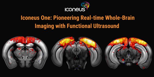
Discover how Iconeus One, the premier functional ultrasound (fUS) system for neuroscientists, is transforming brain function studies. Based on ultrafast plane-wave sonography, Iconeus One of...
FEB 19, 2026 | 8:00 AM
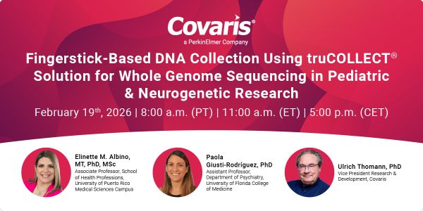
The collection of high-quality genomic DNA remains a major barrier in pediatric and neurodevelopmental research, particularly among children with autism spectrum disorder (ASD) and other neu...
FEB 23, 2026 | 9:00 AM
C.E. CREDITS

Explore decentralized liquid biopsy with SOPHiA GENETICS and MSK-ACCESS®, expanding access, improving outcomes, and accelerating precision oncology....
Loading Comments...